Lab Practical 1
Plant Anatomy
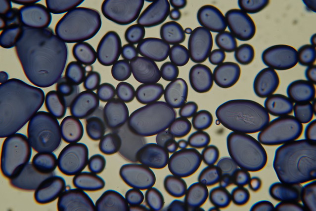
- Photo.
What is this structure?
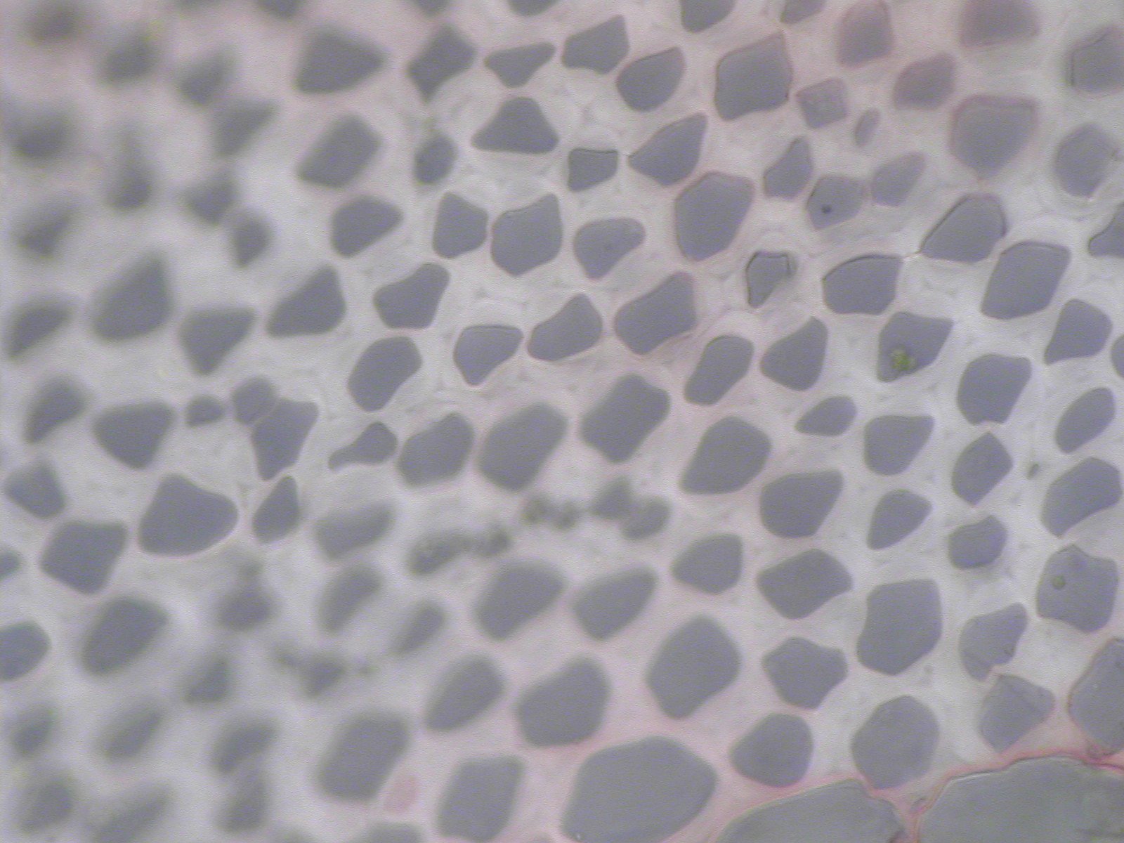
- Photo.
What is this structure?
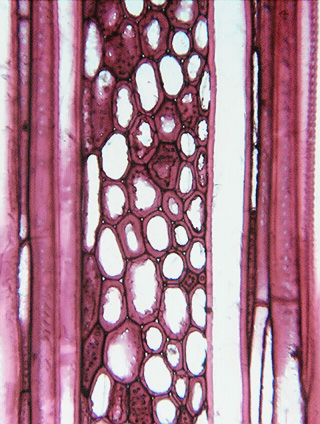
- Photo.
This area shows the junction of three cell walls. What is the thin
line called that cements the cells together?
- Photo.
This Transmission Electron Micrograph shows a mesophyll cell (M) next
to a bundle sheath cell (B) from a corn leaf. Inside this
organelle there are a series of stacked membranes. What are they
called?
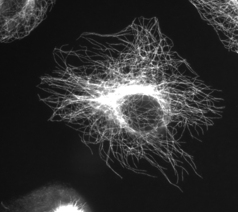
- Photo.
This micrograph shows a cell that has been treated with antibodies to
B-tubulin. What are the structures that are brightly lit up?
- Photo.
This cell, stained with Ioidine Potassium Iodide, shows large ergastic
substances. What are they?
- Photo.
This cell is chuck full of clusters of long, needle-like
structures. What are they?
- Photo.
This is a cross section through the leaf of Ficus.
What is this structure that looks like a mace on a stalk?
- Photo.
This is a section through the seed endosperm of persimmon (Diospyros).
The cell walls are very thick and they serve a function that is unusual
for cell walls. What is that?
- Photo.
This hand section of a stem was stained with phloroglucinol. Why
are the fibers and xylem staining red?
- Photo.
This electron micrograph shows the surface of a plant cell wall with
pores in this area that allow the plasmodesmata of adjacent cells to
connect. From the image, tell whether this is a primary wall or a
secondary wall - and why.
- Photo.
This is a cross section of a Potamogeton
leaf. What is the major cell type seen in the palisade and spongy
mesophyll?
- Photo.
This is a section taken through the leaf of Nymphaea.
What is this tissue called with all the air spaces?
- Photo.
What are these pointy structures called?
- Photo.
This image was taken from the petiole of celery. What types of cells are
shown here with thickened walls?
- Photo.
This is a section from a pear illuminated with fluorescent light.
What types of cells are these?
- Photo.
This is a cross section of the stem of a buttercup (Ranunculus).
What are these cells with thick, red-staining walls?
- Photo.
This is a hand section through the leaf of Peperomia.
What is this darker tissue layer called?
- Photo.
This is a peel made from the leaf of Pelargonium
(geranium). What are these cells called that look like puzzle pieces? BE
SPECIFIC
- Photo.
What are these cells called?
- Photo.
This is a section through an olive (Olea)
leaf. What is this layer here called?
- Photo.
These cells in the palisade mesophyll are full of chloroplasts.
Because of this, specifically what is the cell type?
- Photo.
The guard cells in this plant are bone-shaped (osteoform). What
family do these types of stomata occur?
- Photo.
This slide shows the surface of a leaf with stomata occurring in a row.
The next
slide hows the leaf and a stomata in section. From the
struture, is this plant a monocot, a dicot, or a gymnosperm?
- Photo.
This is a close-up view of the surface of Spanish moss (Tillandsia
usneoides). What are these structures and what is their
function?
- Photo.
This is the water fern Salvinia.
Specifically, what are these structures?
- Photo.
This is a L.S. of the stem of pumpkin (Cucurbita).
Specifically, what is the cell type for these cells?
- Photo.
What terms are used to describe the secondary wall thickenings?
- Photo. In
this slide of Sambucus you can see a progression of cell types.
Specifically, what is this type? Note the walls.
- Photo.
Is this image of a conifer or a dicot?
- Photo.
What is this structure?
- Photo.
Assuming the growth ring at the bottom represents year one, which was
the worst year for this tree in terms of growing conditions?
- Photo.
The cell types above this point are called what and the cell types below
called what.
- Photo.
The majority of cells shown in this image are what type of cell?
- Photo.
What type of section are we seeing here?
- Photo.
Specifically, what is this structure?
- Photo.
Specifically, what are these structures?
- Photo.
What type of section are we seeing here?
- Photo.
What are these square areas called?
- Photo.
Closer view of Sequoia wood,
same region as previous. What type of pit is shown here?
- Photo.
What type of cell is this and does it have a primary or secondary wall?
- Photo.
This is a XS of Catalpa. What
are these structures?
- Photo.
This is a XS of Catalpa. What
term describes the distribution of these structures?
- Photo.
Specifically, what are these structures?
- Photo.
Specifically, what are these structures?
- Photo.
This is a section through Magnolia
wood. Specifically, what are the majority of cells shown here?
- Photo.
This is a tangential section through Maclura
wood. Specifically, what are cells shown here?
- Photo.
This is a radial section through Maclura
wood. Specifically, what type of ray is shown here?
- Photo.
This is a tangential section of a ray, but something has happened to the
ray parenchyma cells. What happened?
- Photo. This
XS of Fraxinus (ash) wood has
parenchyma cells in bands between the vessels. Is it apotracheal or
paratracheal?
Key
1 Nucleus
2 Chloroplast
3 Middle lamella
4 Thylakoids (stack of them called grana)
5 Microtubules
6 Starch
7 Raphides
8 cystolith crystal inside lithocyst
9 nuourishes the seedling upon germination, stores sugar
(mannan)
10 presence of lignin
11 primary - cellulose microfibrils in random array (not
ordered as in secondary wall)
12 parenchyma or chlorenchyma
13 aerenchyma
14 astrosclereids
15 collenchyma (angular)
16 brachysclereids
17 fibers
18 multiple or multiseriate epidermis
19 pavement or subsidiary cells
20 guard cells
21 cuticle
22 chlorenchyma
23 grass (Poaceae)
24 gymnosperm (Pinus)
25 peltate scale or trichome; absorption of water &
nutrients
26 multiseriate trichomes
27 primary xylem - protoxylem
28 annular and helical
29 metaxylem
30 conifer
31 Resin duct
32 year 5
33 early (spring) wood, late (fall) wood
34 tracheids
35 tangential
36 uniseriate ray
37 circular bordered pits
38 radial
39 ray cross fields (with pits)
40 half-bordered pit
41 procumbent ray parenchyma cell with a primary wall
42 vessels
43 ring porous
44 multiseriate rays
45 tyloses
46 fiber-tracheids
47 axial parenchyma
48 heterocellular ray
49 sclerified
50 paratracheal